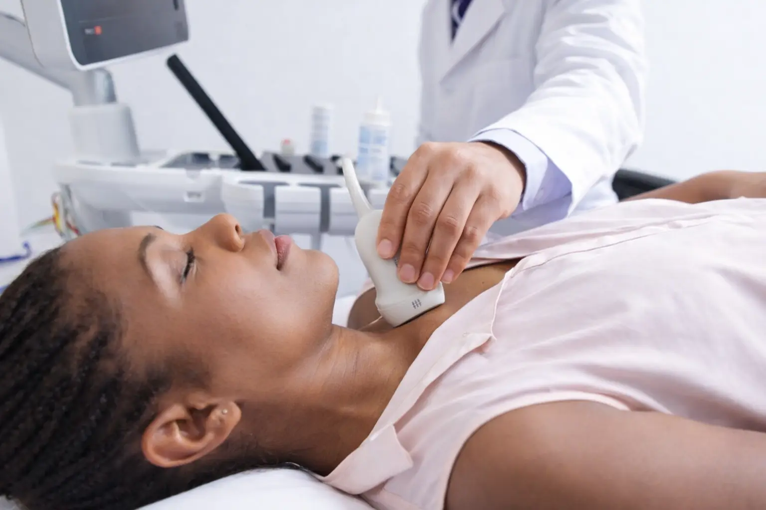
Heart disease remains the leading cause of death among women in the United States, yet women are significantly more likely than men to be underdiagnosed or experience delays in cardiac care. If your doctor has recommended an echocardiogram, you may have questions that go beyond the clinical, questions about privacy, comfort, what you need to remove, and exactly what happens in that room.
This guide answers all of them. Below, you will find a complete, women-focused walkthrough of the echocardiogram procedure, from how to prepare the morning of your appointment to the step-by-step process, special considerations for breast tissue, pregnancy, and implants, and how long it takes to receive your results.
What is an Echocardiogram?
An echocardiogram, often called an echo test, is a non-invasive, ultrasound-based test. It uses high-frequency sound waves to create detailed images of the heart. In addition to imaging the heart, it shows how the heart functions overall. The medical industry uses echocardiograms to assess conditions such as heart failure, valve disease, irregular heartbeat, and blood clots.
Unlike an X-ray or CT scan, an echocardiogram uses no radiation whatsoever. It relies entirely on high-frequency sound waves, the same technology used in pregnancy ultrasounds, making it one of the safest imaging tests available. This is especially important for women who are pregnant or planning to become pregnant.
When performing an echocardiogram on a woman, the procedure is similar to that performed for men. However, it’s essential to keep certain factors in mind due to differences in anatomy and physiology.
Types of Echocardiogram
There are multiple types of heart tests, including cardiac MRI, CT angiography, and stress echocardiograms. These can all be used for different purposes and various conditions. Each type of echocardiogram serves different purposes and is chosen based on symptoms, history, and the required level of detail. Here’s a detailed look depending on the clinical need:
- Transthoracic Echocardiogram (TTE): This is the most commonly performed type, involving placement of the transducer on the chest. It’s used to assess heart function, size, and structure.
- Transesophageal Echocardiogram (TEE): In the TEE test, the transducer is passed through the esophagus to gain clearer images, especially in those with excessive body fat.
- Stress Echocardiogram: This test is performed only when needed. Conducted pre- and post-exercise to assess heart function under strain. It helps identify blockages in the arteries.
- 3D Echocardiogram: This provides an advanced 3D view of the heart’s structure and is beneficial for surgical planning.
- Fetal Echocardiogram: Performed during pregnancy to assess fetal heart development.
Why Do Women Need an Echocardiogram?
Echocardiography is highly helpful in early intervention and treatment of heart-related risks. Women often show cardiovascular symptoms that differ significantly from men. This can lead to symptoms going unnoticed, delaying treatment or resulting in missed diagnoses. Symptoms such as shortness of breath, fatigue, or indigestion-like discomfort may not directly indicate cardiac issues. An echocardiogram helps detect these silent, vague signs of heart disease early. Several other factors can increase the risk of cardiovascular issues in women, including:
- Post-menopause hormonal changes
- Gestational diabetes
- Autoimmune diseases
- Elevated blood glucose levels highlight the critical connection between blood sugar and heart health
How to Prepare for an Echocardiogram as a Woman?
Preparing for a transthoracic echocardiogram (TTE), the most common type, is straightforward. No fasting is required, and no sedation is involved. However, a few practical steps can make the process smoother, particularly for women.
Can You Eat Before an Echocardiogram?
Yes, for a standard transthoracic echocardiogram, you can eat and drink normally before your appointment. There are no dietary restrictions. It is actually advisable to eat a light meal beforehand so you are comfortable during the test. The only exception is a transesophageal echocardiogram (TEE), which requires fasting for about 4–6 hours because a probe is passed through the throat. Your doctor will specify which type of echo you are scheduled for.
Can You Drink Coffee Before an Echocardiogram?
For a standard resting echocardiogram, coffee is generally fine. However, if your appointment includes a stress echocardiogram, you should avoid caffeine for at least 24 hours beforehand, as caffeine can affect your heart rate during the exercise or medication-induced stress portion of the test. When in doubt, follow the instructions provided by the clinician or imaging facility arranging your test.
What to Wear to Your Echocardiogram
Wear a comfortable two-piece outfit: a top that can be easily removed and a separate bottom. You will be given a hospital gown to change into before the test. Avoid one-piece dresses or bodysuits, and skip lotion, powder, or oil on your chest on the day of the test, as these can interfere with gel and probe contact.
What to Bring and Disclose Before the Test
- A list of all current medications, including supplements
- Any prior cardiac test results or imaging reports (if available)
- Pregnancy status, gestational age, or recent pregnancy history
- History of breast augmentation, implants, or chest wall surgery
- History of autoimmune conditions, diabetes, or hypertension
Letting your technician know about breast implants, prior chest surgeries, or pregnancy before the test begins allows them to adjust their approach from the start and ensures clearer imaging results.
Procedure: How is an Echocardiogram Done on a Woman?
The echocardiogram procedure is painless, non-invasive, and typically takes 30 to 60 minutes. Here is exactly what happens during an echocardiogram for women, step by step.
Step 1: Preparation, Privacy, and Gown
When you arrive at the cardiac diagnostic unit, a certified cardiac sonographer or technician will bring you to a private examination room. You will be asked to undress from the waist up and change into a hospital gown that opens at the front.
For women specifically: At the imaging facility, the sonographer or technician should provide a draping sheet to help keep you covered throughout the exam. Only the area of the chest being scanned should be exposed at a time. If you would feel more comfortable with a female technician, ask the facility about this when scheduling your echocardiogram.
If you have a history of breast implants, previous chest surgeries, or are currently pregnant, let your technician know at this stage.
Step 2: Electrode and Sensor Placement
Before imaging begins, small adhesive electrode patches (similar to stickers) are placed on your chest, shoulders, and sides. These electrodes connect to an electrocardiogram (ECG/EKG) monitor that runs simultaneously with the echocardiogram, tracking your heart rhythm in real time throughout the procedure.
For women specifically: The electrode placement is designed to work around the bra line. You will need to remove your bra for proper electrode contact and to allow full probe movement across the chest. The technician will position the draping sheet to keep you comfortable and covered except for the area actively being scanned.
Step 3: Positioning for Optimal Imaging
You will be asked to lie on your left side on the examination table, with your left arm raised above your head. This position causes the heart to shift slightly forward toward the chest wall, making it easier for the transducer to capture clear images.
For women specifically: The left-lateral tilt positioning is particularly important when breast tissue is present, as it helps the heart become more accessible to the probe. Your technician may make small adjustments to your positioning as the scan progresses to capture images from different acoustic windows (angles). This is completely normal and not a sign that anything is wrong.
Step 4: Warm Gel Application and Ultrasound Scanning
A warm, water-based gel is applied to specific areas of your chest. This gel creates a seal between the transducer probe and your skin, eliminating air pockets that would otherwise block sound waves. The technician then gently moves the transducer across your chest, pausing at several standard positions to capture images of your heart from different angles.
For women specifically: In some women, particularly those with larger breast tissue, a higher BMI, or prior chest surgery, the technician may need to gently move or reposition breast tissue to access the best imaging window. This is performed professionally with full draping in place and is a routine part of echocardiography for women. The entire scanning process is not painful; you may feel mild pressure from the probe, but there is no discomfort beyond that.
You may also be asked to hold your breath briefly at certain moments during the scan. This reduces chest movement and results in sharper, more detailed images of specific heart structures.
Step 5: Completion, Gel Removal, and Next Steps
Once all required images have been captured, the gel is wiped away with a soft cloth or tissue. The electrodes are gently removed. You may get dressed immediately and resume all normal activities – driving, eating, working – without any restrictions.
How long does an echocardiogram take?
A standard transthoracic echocardiogram takes 30 to 45 minutes. In some cases, particularly when additional imaging angles are needed due to breast tissue, implants, or body composition, it may take up to 60 minutes. A stress echocardiogram will take longer – typically 60 to 90 minutes – because it includes a pre-test, an exercise or pharmacological stress phase, and a post-stress imaging phase.
After the exam, your images are sent to a qualified physician, often a cardiologist, for interpretation. The clinician who ordered the test or your primary care provider can explain how and when you will receive your results. Timing varies by facility and clinical circumstances.
Special Considerations for Women During an Echocardiogram
Women’s bodies present unique variables that can affect how an echocardiogram is performed and interpreted. Understanding these considerations in advance helps reduce anxiety and ensures you receive the most accurate results possible.
Bra Removal, Breast Exposure, and Modesty
Do you have to remove your bra for an echocardiogram?
Yes. You will be asked to remove your bra before the exam begins. Underwire bras in particular can interfere with electrode placement and prevent the transducer probe from moving freely across the chest. Soft bralettes or sports bras may be loosened or moved rather than fully removed in some cases, depending on your technician’s assessment.
Will your breasts be exposed during the echocardiogram?
Not fully. At no point during the procedure will you be fully undraped. A sheet or gown is used to keep your chest covered at all times, and only the specific area being actively scanned is uncovered at any given moment. The technician will work professionally and efficiently to minimize exposure. If at any point you feel uncomfortable, you have every right to ask for a pause, request a female technician, or ask for additional draping.
Breast Tissue and Larger Chest Size
Breast tissue can create acoustic shadowing that partially blocks ultrasound waves from reaching the heart. This is one of the most common technical challenges in performing echocardiograms on women, and experienced sonographers are trained to address it with specific probe positions and imaging angles called “acoustic windows.”
For women with larger breast tissue or higher body mass, the technician may:
- Gently reposition breast tissue with the patient’s guidance
- Use additional transducer positions below or beside the breast
- Adjust gain and frequency settings on the ultrasound machine
- Request breath-holds at specific moments for sharper image capture
None of these adjustments indicates a problem; they are standard practice for ensuring accurate results in all body types.
Breast Implants and Prior Chest Surgery
Do breast implants interfere with an echocardiogram?
They can create minor imaging challenges. Saline and silicone implants can cast shadows that obscure certain views of the heart, similar to how rib bones naturally shadow some areas. However, echocardiograms can still be performed effectively on women with breast implants. An experienced sonographer will adjust probe angles and use alternative acoustic windows to obtain the images needed. In some cases, 3D echocardiography offers improved visualization when standard 2D views are limited by implants.
Women who have had mastectomy, lumpectomy, or other chest wall surgery should also disclose this prior to the exam, as scarring and tissue changes can similarly affect imaging and require technique adjustments.
Echocardiogram During Pregnancy
Is it safe to have an echocardiogram during pregnancy?
Yes. A transthoracic echocardiogram is completely safe during all stages of pregnancy. It uses no radiation, only sound waves, posing no risk to the mother or the developing baby. Echocardiography during pregnancy may be recommended when a woman has:
- Pre-existing heart disease or a prior cardiac diagnosis
- Gestational hypertension or preeclampsia
- Unexplained shortness of breath, chest pain, or palpitations during pregnancy
- A history of peripartum cardiomyopathy in a previous pregnancy
- Structural heart disease identified during prenatal care
Pregnancy itself causes significant cardiovascular changes, blood volume increases by up to 50%, and cardiac output rises considerably, which is why monitoring maternal heart function is especially important in high-risk pregnancies. If you are pregnant and cardiac monitoring is recommended, your primary care provider can coordinate with your OB-GYN or the appropriate specialist to help ensure you receive the care you need.
High BMI and Body Composition
A higher BMI can make obtaining clear echocardiogram images more technically challenging, as increased chest wall thickness reduces the transmission of sound waves. This does not mean the test cannot be done. Trained sonographers use specialized techniques, including alternative probe positions, harmonic imaging settings, and, in some cases, contrast echocardiography, where a safe contrast agent is injected intravenously to enhance image quality. If standard imaging is insufficient, your provider will discuss alternative or supplementary imaging options with you.
Getting Your Echocardiogram Results: What to Expect
One of the most common questions women ask after their echo test is: how long does it take to get echocardiogram results?
Typical Turnaround Time
After your echocardiogram, a qualified physician, often a cardiologist, reviews and interprets the images. Your ordering clinician or primary care provider can explain the results and discuss any recommended next steps. The time it takes to receive results varies depending on the imaging facility and the clinical circumstances. If the facility identifies an urgent concern, the appropriate medical team will advise you about the next steps.
You will not receive a final interpretation from the sonographer on the day of your test. The technician captures and records the images; the diagnostic interpretation is always performed by a licensed physician.
What Normal Echocardiogram Results Mean
Normal echocardiogram results indicate that your heart’s structure and function fall within healthy ranges. Specifically, a normal echo report will show:
- Normal ejection fraction (typically 55–70%), meaning your heart is pumping blood efficiently
- Normal heart chamber sizes — left and right ventricles and atria within standard measurements
- Normal valve function — no significant regurgitation (leakage) or stenosis (narrowing)
- No pericardial effusion — no abnormal fluid around the heart
- Normal wall motion — heart muscle moving symmetrically and without regional defects
A normal echo is a reassuring finding, but it does not rule out all forms of cardiac disease. If your symptoms persist despite a normal echocardiogram, your physician may recommend additional testing such as a Holter monitor, cardiac stress test, or coronary artery imaging.
What Happens If Results Are Abnormal?
If your echocardiogram reveals an abnormality, such as a reduced ejection fraction, valve dysfunction, or structural defect, your physician will contact you to discuss the findings and outline the next steps in your care plan. An abnormal result does not automatically indicate a serious condition; many findings are manageable with medication, lifestyle changes, or monitoring. Your primary care provider can help explain the findings, discuss what they may mean for your health, and recommend appropriate follow-up care.
Why Should Women Not Delay an Echocardiogram?
Postponing cardiac screening can have unexpected consequences. According to the studies, women are statistically more likely to experience underdiagnosed heart conditions due to the subtlety of symptoms. Any delay in the echocardiogram test can result in the progression of a silent attack and other serious heart conditions.
Key reasons for getting regular cardiac screening include:
- Atypical symptomatology: Symptoms like nausea, fatigue, and anxiety are often dismissed, leading to undetected cardiac conditions.
- Hormonal transitions: Perimenopause and menopause significantly alter lipid profiles and blood pressure.
- Diabetes prevalence: Women with diabetes are at greater risk of heart disease.
- Autoimmune risks: Conditions such as lupus or rheumatoid arthritis disproportionately affect women and elevate cardiovascular risk.
Echocardiography is the best way to assess heart function and for timely intervention. Especially in the presence of diabetes, monitoring the nexus between blood sugar and heart health becomes crucial.
Conclusion
Echocardiography is the ultimate tool to diagnose and treat any heart disease in women. It helps in getting detailed, real-time insight into heart structure and function, helping in both emergency and routine care. By understanding the process and significance of echocardiography, women can benefit from early detection, improved treatment outcomes, and better overall cardiac health.
FAQs
Q1. Do you have to take your bra off for an echocardiogram?
Yes. Underwire bras block electrode placement and probe movement. A gown is provided, and you are kept draped throughout — only the area being scanned is briefly uncovered.
Q2. Will my breasts be exposed during an echocardiogram?
Not fully. A draping sheet covers you at all times. Only the specific chest area being scanned is momentarily uncovered. You can request a female technician when booking.
Q3. Can you eat before an echocardiogram?
Yes — no fasting required for a standard resting echo. Eat and drink normally. The exception is a TEE (transesophageal echocardiogram), which requires 4–6 hours of fasting. Your doctor will confirm which type you are having.
Q4. Can you drink coffee before an echocardiogram?
For a standard resting echo, yes. For a stress echocardiogram, avoid caffeine for 24 hours beforehand as it can interfere with heart rate readings during the stress phase.
Q5. What should you not do before an echocardiogram?
- Do not apply lotion, powder, or oil to your chest
- Avoid caffeine for 24 hours if having a stress echo
- Do not skip disclosing implants, chest surgery, or pregnancy to your technician
- Avoid wearing a one-piece outfit — choose a two-piece for easy access
Q6. How long does an echocardiogram take?
A standard transthoracic echo takes 30 to 45 minutes, occasionally up to 60 minutes if extra imaging angles are needed. A stress echocardiogram takes 60 to 90 minutes.
Q7. How long does it take to get echocardiogram results?
Timing varies by facility and clinical circumstances. The sonographer records images during the test, and a qualified physician interprets them. Your ordering clinician or primary care provider can explain the results and discuss next steps.
Q8. Is an echocardiogram safe during pregnancy?
Yes — completely safe at any stage. It uses only sound waves, no radiation, posing no risk to mother or baby. It is routinely used to monitor maternal heart health in high-risk pregnancies.
Q9. Do breast implants affect an echocardiogram?
They can create minor shadowing over some views, but the test can still be performed effectively. The sonographer adjusts probe angles to work around implants. 3D echocardiography may be used for enhanced clarity when needed.
Q10. Is an echocardiogram painful or uncomfortable?
No. The test is entirely painless. You may feel mild pressure from the probe, but no pain, no needles, and no radiation are involved. The gel is warmed before use for added comfort.
Q11. What is a normal echocardiogram result for a woman?
A normal result shows a healthy ejection fraction (55–70%), normal chamber sizes, proper valve function, and no fluid around the heart. Your physician will review your specific numbers with you.
Q12. Can an echocardiogram detect blocked arteries?
A standard echo does not directly image coronary arteries. However, a stress echocardiogram can detect wall motion abnormalities that suggest reduced blood flow from blockages. For direct artery assessment, a CT angiogram or cardiac catheterization may be recommended alongside your echo.
About the Author